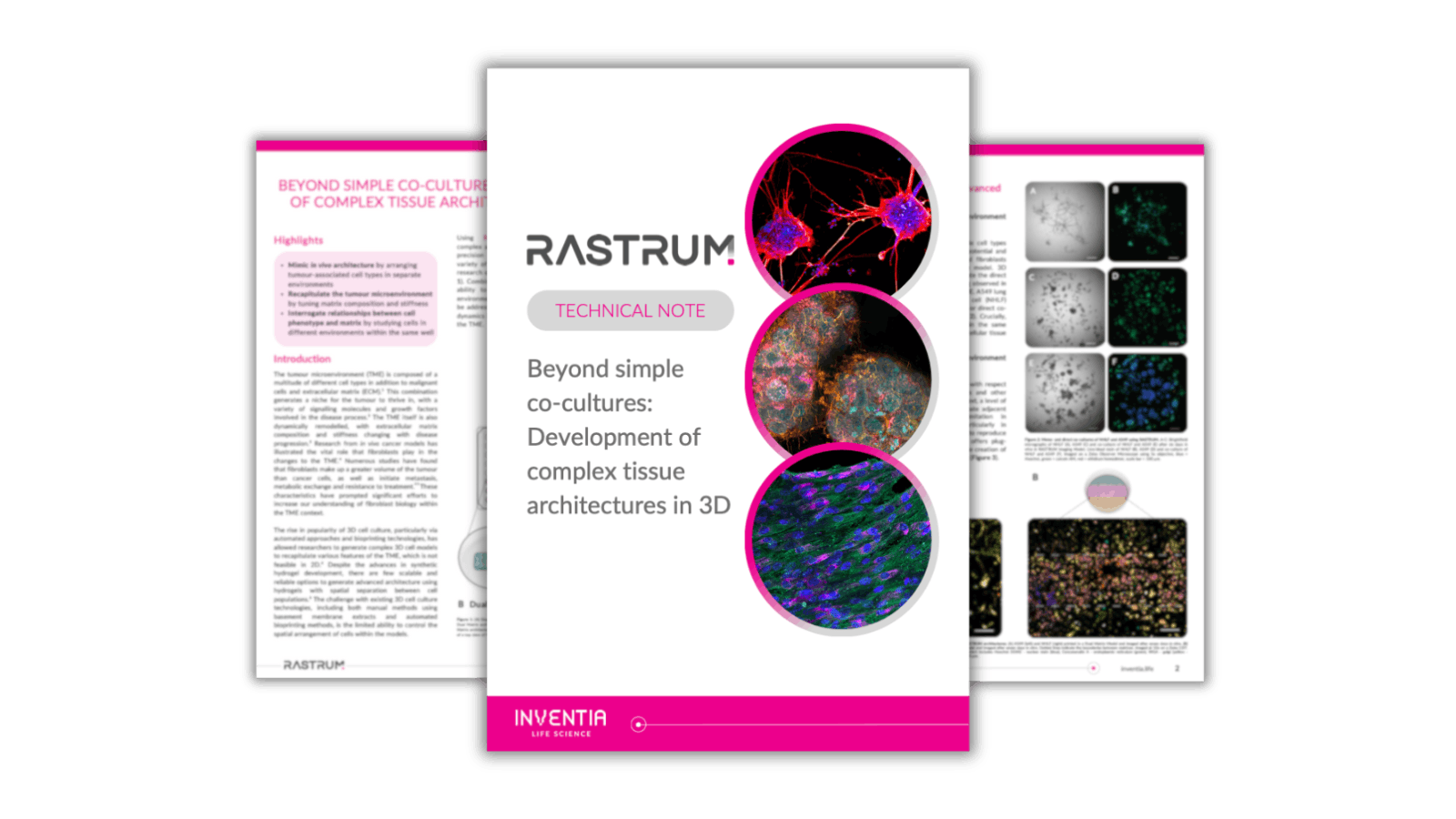
Mimic

Highlights

• Mimic in vivo architecture by arranging tumour-associated cell types in separate environments

• Recapitulate the tumour microenvironment by tuning matrix composition and stiffness

• Interrogate relationships between cell phenotype and matrix by studying cells in different environments within the same well

Technical Note

Want to read the rest of the technical note?

Introduction

The tumour microenvironment (TME) is composed of a multitude of different cell types in addition to malignant cells and extracellular matrix (ECM).¹ This combination generates a niche for the tumour to thrive in, with a variety of signalling molecules and growth factors involved in the disease process.² The TME itself is also dynamically remodelled, with extracellular matrix composition and stiffness changing with disease progression.³ Research from in vivo cancer models has illustrated the vital role that fibroblasts play in the changes to the TME.⁴ Numerous studies have found that fibroblasts make up a greater volume of the tumour than cancer cells, as well as initiate metastasis, metabolic exchange and resistance to treatment.⁵ These characteristics have prompted significant efforts to increase our understanding of fibroblast biology within the TME context.

Tech Note

The rise in popularity of 3D cell culture, particularly via automated approaches and bioprinting technologies, has allowed researchers to generate complex 3D cell models to recapitulate various features of the TME, which is not feasible in 2D.⁶ Despite the advances in synthetic hydrogel development, there are few scalable and reliable options to generate advanced architecture using hydrogels with spatial separation between cell populations.⁶ The challenge with existing 3D cell culture technologies, including both manual methods using basement membrane extracts and automated bioprinting methods, is the limited ability to control the spatial arrangement of cells within the models.

Using RASTRUM drop-on-demand technology, complex architectures are now possible due to high-precision droplet deposition. RASTRUM provides a variety of different architectures tailored to specific research questions about cancer pathogenesis (Figure 1). Combined with the tunable RASTRUM Matrix, the ability to create distinct, spatially separated cell environments allows complex research questions to be addressed, facilitating better understanding of the dynamics between cancer cells and fibroblasts within the TME.

Technical Note

Want to read the rest of the technical note?

References

  1. Zanoni M., Piccinini F., Arienti C. et al. 3D tumor spheroid models for in vitro therapeutic screening: a systematic approach to enhance the biological relevance of data obtained. Sci Rep. 2016; 6, 19103 doi:10.1038/srep19103
  2. Murakami T., Hiroshima Y., Matsuyama R. et al. Role of the tumor microenvironment in pancreatic cancer. Ann Gastroenterol Surg. 2019; 3(2):130-137, doi:10.1002/ags3.12225
  3. Sharbeen G., McCarroll J.A., Akerman A. et al. Cancer- Associated Fibroblasts in Pancreatic Ductal Adenocarcinoma Determine Response to SLC7A11 Inhibition. Cancer Res, 2021; 81(13):3461-3479, doi:10.1158/0008-5472.CAN-20-2496
  4. Xing F., Saidou J., Watabe K. Cancer associated fibroblasts (CAFs) in tumor microenvironment. Front Biosci (Landmark Ed). 2010; 15(1):166-79, doi:10.2741/3613
  5. Hu D., Li Z., Zheng B. et al., Cancer-associated fibroblasts in breast cancer: Challenges and opportunities. Cancer Commun (Lond). 2022; 42(5):401-434, doi:10.1002/cac2.12291
  6. Kapałczyńska M., Kolenda T., Przybyła W. et al., 2D and 3D cell cultures - a comparison of different types of cancer cell cultures. Arch Med Sci. 2018; 14(4): 910–919, doi:10.5114/aoms.2016.63743