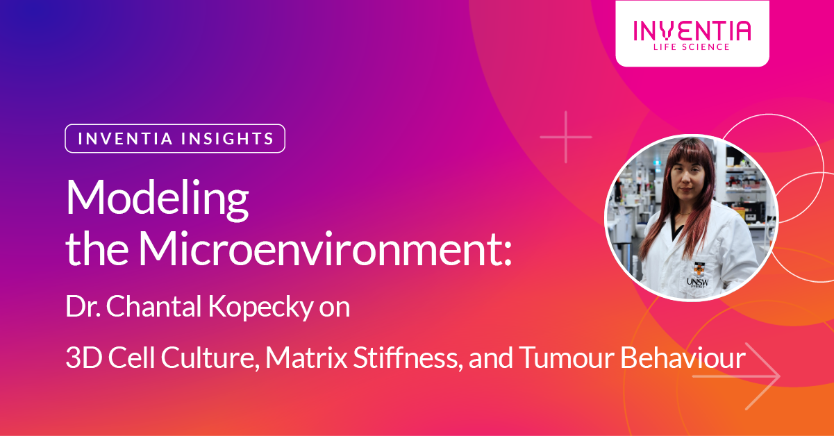
Modeling the Microenvironment: Dr. Chantal Kopecky on 3D Cell Culture, Matrix Stiffness, and Tumour Behaviour
For Chantal Kopecky, Ph.D., of the University of New South Wales (UNSW Sydney), understanding cancer requires more than just looking at the cells—it demands recreating the context in which they live. As a Postdoctoral Research Fellow at UNSW working in Prof. Kris Kilian’s biomaterials and matrix engineering lab, Chantal is using cutting-edge technologies to model the tumour microenvironment and decode how physical cues like matrix stiffness shape cancer progression and metastasis.
With a background in cancer biology and a growing toolkit of bioengineered 3D systems, her research spans melanoma and breast cancer, bridging the gap between mechanobiology and translational oncology.
We caught up with Chantal to learn more about her work—and how platforms like RASTRUM™ Allegro are accelerating her ability to explore tumour heterogeneity, cell plasticity, and personalized response to therapy.
From biology to bioengineering
Chantal’s journey into matrix engineering was rooted in a desire to bridge scientific disciplines. With her training in cancer biology, she was drawn to the translational potential of understanding how physical and mechanical forces shape tumour behaviour—something traditional 2D culture models often fail to capture.
"I'm a cancer biologist, and for the past four years I've been working within a biomaterials and matrix engineering group at UNSW Sydney led by Prof Kris Kilian. My research focuses on applying advanced biomaterials to model the tumour microenvironment, with the goal of better understanding how biomechanical cues within that environment influence cancer progression and metastasis.
Specifically, I work with melanoma and breast cancer models, aiming to capture the complexity of the tumour microenvironment in a way that's both accurate and reproducible. By creating bioengineered matrices with tuneable mechanical properties, I'm able to study how changes in the matrix—such as stiffness or architecture—affect cancer cell plasticity and phenotype.
What really drew me to this field is the intersection of biology, engineering, and translational science. Understanding the physical context of tumours opens up new possibilities for targeted therapies and more predictive in vitro models, which I find incredibly exciting and impactful."
Rethinking the role of matrix stiffness
In recent years, a prevailing narrative in cancer biology has focused on how stiff microenvironments promote tumour progression. But Chantal’s research, using both 2D and 3D systems, has begun to reveal a more nuanced story—one where softness may also play an active and dynamic role.
"One of the most exciting findings from our study was the distinct effect of soft matrices on cancer cell behaviour and phenotype across different cancer types. Traditionally, much of the literature has focused on how stiff tumour microenvironments promote cancer cell migration, invasion, and progression. However, using both 2D and 3D models, we found that softer matrices can also drive significant and specific changes in cancer cell plasticity and even drug sensitivity.
These soft environments seem to induce a unique cellular response that’s different from what we see with stiffer conditions—suggesting that the mechanical cues in the microenvironment don’t just act as a binary switch but rather influence a spectrum of behaviours. This has important implications for how we understand tumour heterogeneity and for the development of more physiologically relevant models and therapeutic strategies."
Why 3D matters
This discovery wouldn’t have been possible without moving beyond the limitations of 2D cell culture. As Chantal explains, while 2D systems serve an important function, they simply can’t replicate the spatial or mechanical complexity that real tumours experience.
"While traditional 2D cell culture models are useful for initial testing—they're quick, cost-effective, and relatively easy to perform—they fall short in capturing the complexity, spatial organization, and dynamic interactions found in real tissues. In contrast, 3D cell culture systems provide a much more physiologically relevant context, allowing us to more accurately mimic the tumour microenvironment.
In our work, this has been critical since the dimensionality and tuneable properties of our 3D biomaterial platforms allow us to model not just biochemical signals, but also mechanical and architectural cues that influence cancer cell behaviour. We've observed that while 3D models often help validate findings from 2D systems, there are notable cases where cell responses diverge completely between the two settings. This can include differences in morphology, drug sensitivity, and cell–matrix interactions for example.
These discrepancies highlight the need for advanced 3D models to generate data that are more predictive of in vivo outcomes. Especially when studying complex processes like cancer metastasis and plasticity, leveraging 3D systems is essential for capturing the nuance of how tumour cells interact with their surrounding environment."
Accelerating insights with RASTRUM
Enter RASTRUM Allegro. For Chantal, the platform has provided a way to scale complexity—making it possible to run high-throughput experiments while retaining fine control over mechanical cues like matrix stiffness.
"In addition to the general advantages of using 3D models for more physiologically relevant research, the RASTRUM system has significantly advanced our ability to generate these models at a high-throughput scale. One of the key strengths of RASTRUM, for me, is its flexibility — being able to select from a wide range of matrix compositions, incorporating different peptides and stiffness levels, allows for highly customizable model systems tailored to specific research questions.
For my work, this has been particularly valuable allowing me to use matrix stiffness as a central feature in modelling the tumour microenvironment, and the platform's high-throughput capability has enabled me to print multiple microplates with different conditions in parallel. This enables systematic investigations of how specific matrix properties influence cancer cell behaviour — with side-by-side comparisons within a single cancer type, and flexibility to adapt the system for use with different cancers, such as melanoma and breast cancer.
The ability to ‘mix and match’ conditions, while maintaining consistency and reproducibility, really sets the RASTRUM platform apart. It’s a powerful tool that’s helped streamline complex experiments and accelerate the pace at which we can generate and test meaningful hypotheses."
Next steps: Toward personalized cancer models
Looking forward, Chantal’s vision is to take these findings one step closer to the clinic—by working with patient-derived samples and ultimately building out a pipeline for personalized therapeutic screening.
"We’ve generated some really intriguing data using the RASTRUM bioprinted cancer models, particularly in terms of phenotypic profiling and differential drug responses. These findings highlight the potential of our system not only for understanding cancer biology in a more physiologically relevant way but also for identifying context-dependent therapeutic responses.
So far, we’ve been working primarily with commercial melanoma and breast cancer cell lines. As a next step, we’re planning to transition to patient-derived cell lines to better capture tumour heterogeneity and improve clinical relevance. This would allow us to move toward, e.g. setting up a personalized drug screening platform that could ultimately help inform treatment strategies and contribute to more tailored therapeutic approaches.
Looking ahead, I see this work evolving into a robust pipeline for high-throughput testing in engineered microenvironments — bridging the gap between basic cancer biology, biomaterials science, and translational oncology."
Advice for researchers making the 3D leap
Making the move from 2D to 3D culture can feel daunting, but Chantal encourages researchers to take the first step, even if it means starting small or seeking out collaboration.
"I would absolutely encourage researchers to make the transition from 2D to 3D cell culture. While 2D models are valuable for initial screening, 3D systems offer a much more physiologically relevant context that can reveal nuances in cell behaviour, drug response, and tissue interactions that simply aren’t captured in 2D.
If you're hesitant, a great way to start could be by collaborating with a group already working with 3D systems. This allows you to get hands-on experience without needing to invest heavily upfront. Alternatively, reaching out to companies that offer 3D culture platforms — many may provide starter kits, training, or technical support — may be a very accessible entry point.
Ultimately, moving to 3D culture doesn't mean abandoning everything you’ve done in 2D; it’s about enhancing your model to ask deeper, more translationally relevant questions. The insights gained from a 3D context are well worth the learning curve."
Want to learn more about how researchers are using RASTRUM to explore tumour biology, drug response, and translational oncology?
Explore our Cancer Applications page →